Back pain can be a perplexing and frustrating experience, especially when it seems to come out of nowhere and refuses to go away. Among the myriad causes of back discomfort, herniated discs stand out as a particularly challenging condition, often shrouded in mystery and complexity. These elusive culprits can cause symptoms ranging from sharp pain to numbness or weakness in the limbs, making everyday activities difficult and uncomfortable.
A herniated disc occurs when the soft, jelly-like center of a spinal disc pushes through a crack in its tougher exterior casing. This can irritate nearby nerves, leading to the aforementioned symptoms. While some people may experience excruciating pain, others might only feel a slight discomfort or even no symptoms at all. This variability can make diagnosing a herniated disc a tricky endeavor.
The role of MRI in diagnosing herniated discs
Enter the MRI, or magnetic resonance imaging, a powerful tool in the diagnostic arsenal for uncovering the secrets of herniated discs. Unlike other imaging methods such as X-rays or CT scans, an MRI is non-invasive and does not involve exposure to harmful radiation. This makes it a preferred choice for doctors aiming to get a clear picture of what’s happening inside the spine. The MRI can provide highly detailed images of the spinal discs, nerves, and surrounding structures, allowing for a precise diagnosis.
The purpose of this post is to shed light on the crucial role that MRIs play in diagnosing herniated discs. We aim to demystify the process, explaining how this technology works and what patients can expect from their results. Whether you're experiencing symptoms and considering an MRI or simply curious about the process, this post will guide you through the essentials of how MRIs reveal the hidden truths of herniated disc conditions.
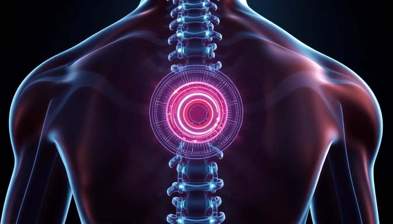
understanding MRI technology for herniated discs
Magnetic Resonance Imaging, or MRI, stands as the gold standard for evaluating spinal disc injuries, including herniated discs. With an impressive accuracy rate of 95%, this technology offers unparalleled insights into the spine's condition. Unlike X-rays or CT scans, MRIs provide detailed images with a spatial resolution as fine as 0.5mm, allowing healthcare professionals to identify even the smallest disc abnormalities.
The absence of harmful radiation is another significant advantage of MRI technology. This makes it a safer option for patients who require detailed imaging of their spine. The MRI uses powerful magnets and radio waves to create detailed images of the body's internal structures, offering a comprehensive view that other imaging techniques cannot match. This detail is crucial when planning treatment, as it allows for precise targeting of the problem area.
when is an MRI necessary for a herniated disc?
While not every case of back pain necessitates an MRI, certain scenarios do call for this advanced diagnostic tool. If a patient experiences persistent symptoms despite undergoing conservative treatments like physical therapy or medication, an MRI may be recommended. Additionally, if neurological symptoms such as numbness, tingling, or muscle weakness worsen, an MRI becomes crucial to assess the extent of nerve involvement.
In situations where surgery is being considered as a treatment option, an MRI provides essential information to guide surgical planning. By revealing the exact location and severity of the herniation, an MRI helps surgeons determine the most appropriate surgical approach, if necessary.
interpreting MRI results for herniated discs
Reading MRI results requires a trained eye, as the images reveal complex structures within the spine. Doctors look for signs of herniation, where the disc material has protruded beyond its normal boundary. On an MRI, a herniated disc appears as a bulging area that may compress nearby nerves, leading to the symptoms experienced by the patient.
It's important to differentiate between herniated, bulging, and ruptured discs, as each presents differently on an MRI. A bulging disc shows a more uniform protrusion, while a ruptured disc indicates that the disc material has broken through its outer layer. These distinctions are vital for developing an effective treatment plan, as they dictate the severity and urgency of the intervention needed.
Understanding the implications of MRI findings is crucial for planning the next steps in treatment. For many patients, conservative treatments may suffice, but for others, the MRI results may indicate a need for more aggressive interventions. This could range from injection therapies to surgical options, depending on the severity of the herniation and the patient's overall health.
In conclusion, MRIs play an indispensable role in the diagnosis and management of herniated discs. Their ability to provide detailed, accurate images without the risk of radiation makes them an invaluable tool in modern medicine. By understanding when an MRI is necessary and how to interpret the results, patients and healthcare providers can work together to develop a personalized treatment plan that addresses the root cause of back pain and improves quality of life.
Treatment options following an MRI diagnosis
Once a herniated disc has been diagnosed through an MRI, the focus shifts to exploring the most effective treatment options. Fortunately, the majority of cases can be managed with conservative approaches, allowing patients to return to their daily activities without the need for invasive procedures.
Conservative approaches
For many individuals diagnosed with a herniated disc, medications and physical therapy are the first line of treatment. Over-the-counter pain relievers and anti-inflammatory medications can help alleviate discomfort, while physical therapy focuses on strengthening the muscles supporting the spine. This approach is effective in approximately 9 out of 10 cases, with most patients experiencing significant improvement without the need for surgery.
Injection therapies
In cases where conservative treatments do not provide sufficient relief, injection therapies may be considered. Selective nerve root blocks (SNRBs) and epidural steroid injections (ESIs) are two common options. SNRBs have a success rate of around 75%, while ESIs can offer relief in 76-88% of cases. These injections work by delivering anti-inflammatory medication directly to the affected area, reducing swelling and alleviating pain.
Surgical options
For patients who do not respond to conservative or injection therapies, surgical intervention may be necessary. Surgical options for herniated discs include procedures such as discectomy or microdiscectomy, which involve removing the portion of the disc that is pressing on the nerve. Recovery timelines for these surgeries typically range from 2 to 3 months, with most patients experiencing a substantial reduction in symptoms.
Conclusion
In conclusion, MRIs are a critical tool in the accurate diagnosis and management of herniated discs. By providing detailed images of the spine, MRIs allow healthcare professionals to develop targeted treatment plans tailored to each patient's needs. From conservative approaches to surgical options, understanding the full spectrum of treatments available ensures that patients receive the most effective care possible.
Additionally, incorporating ergonomic solutions into the recovery process can further enhance outcomes. Ergonomic products, such as supportive chairs and adjustable desks, can help prevent future disc issues and support long-term spinal health. At Anodyne, we are committed to providing solutions that promote recovery and prevent recurrence, ensuring our clients maintain a healthy, active lifestyle.
Frequently Asked Questions
What is a herniated disc, and how does it differ from a bulging disc?
A herniated disc occurs when the soft inner material of a spinal disc pushes through a crack in its outer casing, potentially irritating nearby nerves. In contrast, a bulging disc involves the disc protruding outward without the inner material breaking through. Both conditions can cause similar symptoms but differ in severity and treatment approaches.
How does an MRI work, and why is it preferred for diagnosing herniated discs?
An MRI uses powerful magnets and radio waves to create detailed images of the body's internal structures. It is preferred for diagnosing herniated discs due to its high accuracy and ability to provide clear images without exposing patients to radiation, unlike X-rays or CT scans.
What should I expect during an MRI scan for a herniated disc?
During an MRI scan, you will lie still on a table that slides into the MRI machine. The procedure is painless and typically lasts between 30 to 60 minutes. You may hear loud noises from the machine, but earplugs or headphones can help minimize the sound. It's important to remain still to ensure clear images are captured.
How are MRI results interpreted, and what do they mean for my treatment?
MRI results are interpreted by trained radiologists and medical professionals who look for signs of disc herniation, such as protrusion or nerve compression. These findings guide treatment decisions, helping determine whether conservative methods, injections, or surgery are the best course of action.
Are there any risks associated with MRI scans?
MRIs are considered safe and non-invasive, with no exposure to harmful radiation. However, individuals with certain implants or metal in their body should inform their doctor, as these may interfere with the MRI's magnetic field. Otherwise, the procedure poses minimal risk to patients.
Sources
- Mayo Clinic Staff. (2023). "Herniated Disk: Diagnosis & Treatment." Mayo Clinic.
- Deuk Spine Institute. (2023). "Herniated Disc Diagnosis and Treatment."
- Cleveland Clinic. (2023). "Herniated Disc: Symptoms, Causes, and Treatment." Cleveland Clinic.
- Weill Cornell Medicine. (2023). "Herniated Disc." Weill Cornell Brain and Spine Center.
- Pauza, K. (2023). "Understanding Herniated Discs." Dr. Kevin Pauza.
- Sciatica.com. (2023). "Herniated Disc Treatment Options."
- Barrow Neurological Institute. (2023). "Herniated Disc: Overview and Treatment."