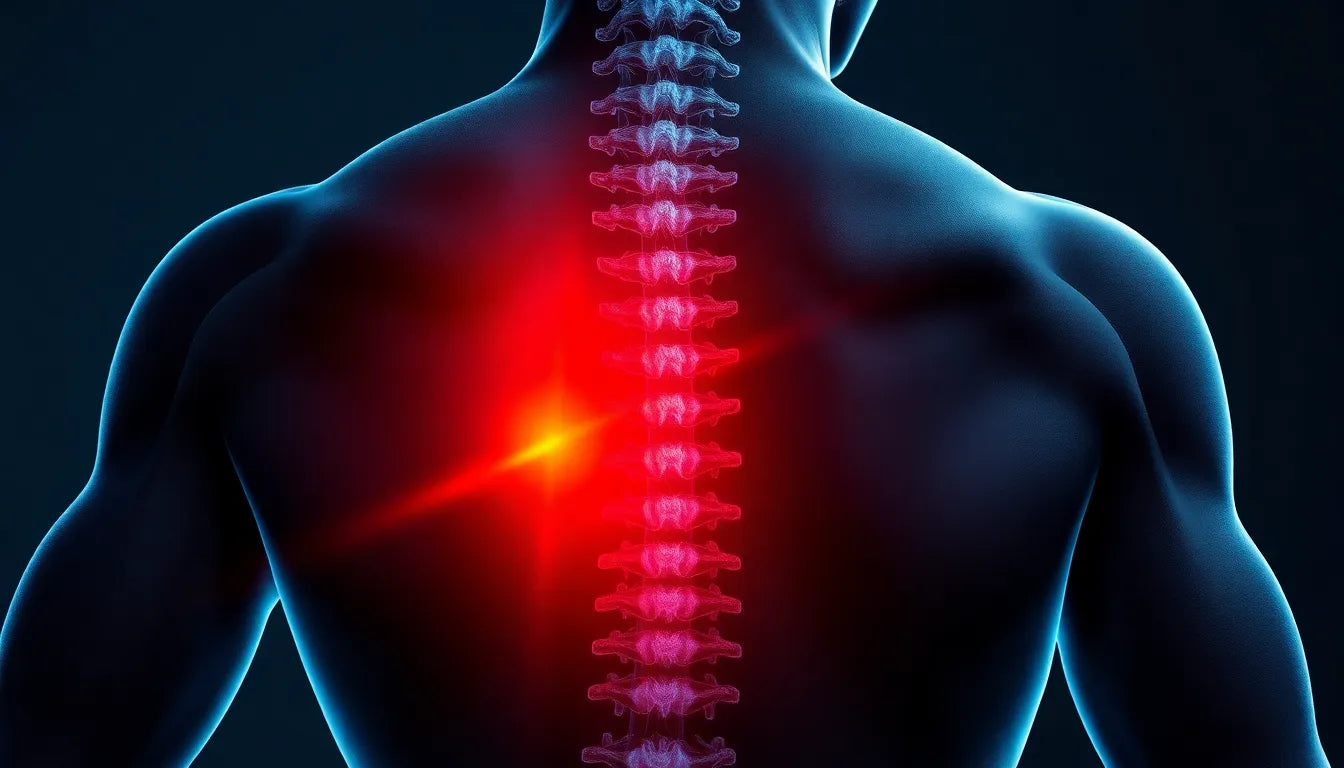
Understanding what a herniated disc looks like is crucial for individuals seeking to comprehend their spinal health. A herniated disc, often referred to as a slipped or ruptured disc, occurs when the soft inner gel of a spinal disc protrudes through its tougher outer layer. This condition can lead to significant discomfort and impact overall spinal function. The spine, a vital component of our body's structure, relies on healthy discs to absorb shock and allow for flexible movement. When a disc becomes herniated, it can press on nearby nerves, leading to pain, numbness, or weakness in various parts of the body.
The role of imaging in diagnosing herniated discs
Imaging plays a pivotal role in diagnosing and understanding herniated discs. For many, the journey begins with an MRI or magnetic resonance imaging scan, which is the gold standard for visualizing these spinal issues. Unlike X-rays, which primarily show bones, MRIs provide detailed images of soft tissues, including discs, nerves, and the spinal cord. This detailed view is essential for accurately diagnosing a herniated disc and understanding its severity and potential impact on the spine and surrounding nerves.
Individuals often seek out images of herniated discs to better interpret their MRI results or to gain a clearer understanding of their diagnosis. This search is driven by the need to visualize the problem and comprehend its implications on their health. Understanding what a herniated disc looks like through imaging can empower patients to make informed decisions about their treatment and manage their expectations regarding recovery and lifestyle adjustments.
Visualizing the problem with MRI
Magnetic resonance imaging is unparalleled in its ability to reveal the nuances of spinal health, particularly when it comes to herniated discs. On an MRI scan, a herniated disc typically appears as a bulge or protrusion extending beyond the normal boundaries of the disc. This protrusion can impinge on adjacent nerve roots, which is often the source of symptoms like pain, tingling, or numbness. By visualizing the herniated disc, patients and healthcare providers can better understand the anatomical changes occurring within the spine.
Seeing the problem is a crucial step in comprehending its implications. MRI images not only confirm the presence of a herniated disc but also provide insights into the extent of the condition. This visual evidence allows for a tailored approach to treatment, whether it involves physical therapy, medication, or in some cases, surgical intervention. Understanding the visual characteristics of a herniated disc is a vital part of the diagnostic process, offering clarity and direction for both patients and practitioners.
mri as the diagnostic tool for herniated discs
Magnetic resonance imaging, or MRI, is the cornerstone for diagnosing herniated discs. This advanced imaging technique offers unparalleled insights into the spinal structure, allowing for a clear depiction of disc abnormalities. When examining an MRI, a herniated disc is typically visible as a bulge or protrusion that extends beyond the normal confines of the disc space. This protrusion can be seen pressing against the spinal canal or nerve roots, which is often the underlying cause of associated symptoms.
Understanding the different MRI views is crucial for accurate interpretation. The axial view, which provides a cross-sectional image, is particularly useful for identifying the extent of disc herniation and its impact on surrounding structures. In contrast, the sagittal view offers a side perspective, showcasing the disc's vertical alignment and any displacement. Together, these views offer a comprehensive understanding of the herniated disc's characteristics and its potential effects on the spine.
using visual aids to enhance understanding
Visual aids, such as labeled diagrams and annotated MRI images, play a vital role in illustrating the presence and implications of a herniated disc. These tools highlight the herniated areas, often marked with circles or arrows, making it easier for patients to identify the problematic regions. By visually pinpointing nerve compression and disc degeneration, these aids enhance comprehension and facilitate more informed discussions between patients and healthcare providers.
For instance, a comparison between healthy and herniated discs can vividly demonstrate the structural differences. In a healthy disc, the nucleus pulposus is contained within the annulus fibrosus, maintaining a smooth and even contour. However, in a herniated disc, the nucleus protrudes, disrupting this contour and potentially compressing nearby nerves. Such visual comparisons are instrumental in helping patients grasp the anatomical changes and their potential impact on their symptoms.
clinical explanation and symptom associations
The structural changes seen in herniated discs are closely linked to various symptoms. Commonly, individuals with a herniated disc experience pain, tingling, or numbness, often radiating along the path of the affected nerve. This is due to the pressure exerted by the herniated disc on the nerve roots, which can lead to irritation and inflammation. Understanding these associations is crucial for both diagnosing the condition and developing an effective treatment plan.
Moreover, herniated discs pose potential risks of further degeneration or nerve damage if left untreated. Recognizing these risks early through imaging and clinical evaluation can prevent complications and guide appropriate interventions. Educational guides, which often use slideshows or visual aids, are valuable resources for patients. They bridge the gap between complex medical information and patient understanding, providing clear explanations of how their symptoms are related to the visual evidence seen in MRI scans.
educational resources for patient empowerment
Access to educational resources is essential for empowering patients in their healthcare journey. By understanding MRI scans and the correlation between their symptoms and the visual evidence, patients can make more informed decisions about their treatment options. Resources that offer detailed explanations, such as those provided by healthcare professionals or reputable medical websites, equip patients with the knowledge needed to actively participate in their care.
These resources often include visual guides that explain the patterns of radiating pain and symptom progression, offering insights into the underlying causes of discomfort. By demystifying the diagnostic process and clarifying the implications of a herniated disc, these guides foster a sense of reassurance and confidence in patients, enabling them to engage more fully in discussions about their treatment and recovery plans.
for practitioners and patients: bridging the gap
Understanding herniated disc images is not only crucial for patients but also for healthcare practitioners. For medical professionals, the insights gained from MRI scans are complemented by anatomical and clinical examinations. These examinations help identify which specific nerve is affected by the herniation, providing a comprehensive view that supports MRI findings. This holistic approach ensures that practitioners can tailor treatment plans to address both the structural and symptomatic aspects of the condition.
For patients, the journey through understanding herniated disc images can be daunting. However, clarity and reassurance are key elements in guiding them through their scans. By demystifying the images and explaining their implications, healthcare providers can empower patients to make informed decisions about their treatment. Understanding these images helps patients grasp the connection between their symptoms and the underlying anatomical changes, influencing their treatment choices and expectations.
frequently asked questions
How is a herniated disc diagnosed?
A herniated disc is typically diagnosed through a combination of physical examinations and imaging techniques. During a physical exam, a healthcare provider will assess your symptoms, reflexes, and muscle strength. An MRI scan is often used to confirm the diagnosis, as it provides detailed images of the spinal discs and can clearly show any herniation or nerve compression.
What are the symptoms of a herniated disc?
The symptoms of a herniated disc can vary depending on the location and severity of the herniation. Common symptoms include pain, tingling, or numbness in the affected area, which may radiate to the arms or legs. Muscle weakness and difficulty in movement may also occur, as the herniated disc can press on nearby nerves.
Can herniated discs heal on their own?
In some cases, herniated discs can heal on their own over time as the body reabsorbs the protruding material. However, this process can take weeks to months, and not all herniated discs will resolve without intervention. It is important to consult with a healthcare provider to determine the best course of action, which may include physical therapy, medication, or other treatments.
What treatments are available for herniated discs?
Treatment options for herniated discs range from conservative approaches to surgical interventions. Conservative treatments include physical therapy, pain management with medications, and lifestyle modifications. If these methods are ineffective, or if the herniation is severe, surgical options such as discectomy or spinal fusion may be considered to relieve pressure on the nerves and stabilize the spine.
conclusion
Understanding herniated disc images is a vital component of both diagnosis and treatment. By combining visual aids with clear explanations, healthcare providers can empower patients to engage actively in their healthcare journey. Recognizing the visual characteristics of a herniated disc not only aids in accurate diagnosis but also informs treatment decisions, ultimately enhancing patient outcomes and quality of life.
Sources
- ADR Spine. "Understanding Herniated Discs with MRI."
- WebMD. "Understanding Herniated Discs: Basics and Symptoms."
- Austin Spine Health. "Interpreting MRI Results for Herniated Discs."
- iStock. "Herniated Disc Images and Diagrams."
- Physiopedia. "Clinical Examination of Herniated Discs."
- Deuk Spine. "A Comprehensive Guide to Herniated Discs."