Herniated discs are a common source of pain and discomfort, affecting countless individuals worldwide. Despite their prevalence, there remains a significant amount of confusion and misunderstanding about what a herniated disc actually looks like. By delving into the visual aspects of this condition, we can demystify it and empower those affected with knowledge that aids in understanding their symptoms and treatment options.
why understanding herniated discs is important
Many people harbor misconceptions about herniated discs, often imagining them as dramatic, catastrophic events. In reality, the appearance and impact of a herniated disc can vary greatly, ranging from minor bulges to significant protrusions that press on nearby nerves. Understanding what a herniated disc looks like is crucial for both patients and healthcare providers to accurately diagnose and treat the condition. This post aims to illuminate the visual characteristics of herniated discs, using a blend of technical insights and accessible language to cater to a wide audience.
the role of visual aids in understanding medical conditions
Visual aids, such as MRI and CT scans, play a pivotal role in comprehending medical conditions like herniated discs. These imaging techniques provide a detailed look at the spine's structure, revealing the precise nature and extent of disc herniation. For patients experiencing symptoms like back pain or sciatica, seeing what a herniated disc looks like can be both enlightening and reassuring. It bridges the gap between clinical symptoms and their anatomical causes, offering clarity and aiding in effective communication with healthcare professionals.
introduction to herniated discs
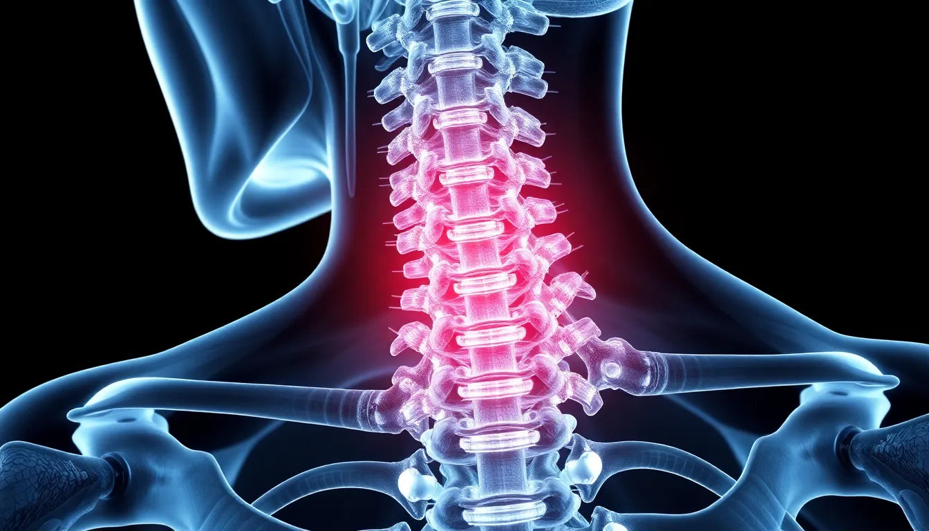
Herniated discs occur when the soft, gel-like center of a spinal disc pushes through a tear in its tougher exterior casing. This can happen anywhere along the spine, but it is most common in the lower back. The herniation can lead to pain, numbness, or weakness in the limbs if it compresses nearby nerves. By exploring the visual representation of herniated discs, readers will gain insights into both the medical imaging and the symptoms associated with this condition, setting the stage for a deeper understanding in the sections that follow.
understanding herniated discs through imaging
When it comes to diagnosing a herniated disc, medical imaging is an invaluable tool. MRI (Magnetic Resonance Imaging) and CT (Computed Tomography) scans are the most commonly used methods to visualize herniated discs, offering detailed images of spinal structures. These scans are crucial for identifying the exact location and severity of the herniation, which in turn informs treatment decisions.
MRIs are particularly effective in highlighting the soft tissues of the spine, including the discs and surrounding nerves. They provide high-resolution images that can reveal even subtle changes in disc structure. On an MRI, herniated discs often appear as bulges or protrusions of the disc material. The signal intensities on T1-weighted (T1W) and T2-weighted (T2W) images can help radiologists differentiate between normal and abnormal disc conditions. T2W images, in particular, are adept at showing the presence of fluid, which can indicate inflammation around the herniated area.
CT scans, while less detailed in soft tissue contrast compared to MRIs, are still useful in visualizing the bony structures of the spine and can help in assessing the extent of disc herniation and any associated bone changes. They are often used when MRI is not an option due to patient contraindications or other factors.
different planes and views in imaging
To fully understand the extent of a herniated disc, viewing the spine in different planes is essential. The sagittal plane provides a side view of the spine, allowing for a clear visualization of the spinal cord and the alignment of the vertebrae. This view is particularly useful in assessing the degree of disc protrusion and its impact on the spinal canal.
The axial plane, on the other hand, offers a cross-sectional view of the spine. This perspective is crucial for evaluating the degree of nerve compression and the disc's impact on adjacent structures. By examining both sagittal and axial views, healthcare providers can gain a comprehensive understanding of the herniation's characteristics and its potential to cause symptoms.
simplified explanation for patients
For patients, understanding the complex images produced by MRI and CT scans can be daunting. In simple terms, a herniated disc can be thought of as a "slipped" or "ruptured" disc that disrupts the normal alignment of the spine. This misalignment can lead to the disc pressing on nearby nerve roots, causing pain, numbness, or weakness in the affected area.
The clarity of MRI images plays a significant role in accurately diagnosing the condition and understanding its implications. By visualizing the herniation, patients can better grasp the source of their symptoms and the rationale behind their treatment plan.
the role of video and animated content
In addition to static images, animations and videos can be powerful tools for illustrating how herniated discs affect the spine. Platforms like Spine Health use dynamic visuals to demonstrate the process of disc herniation and its impact on spinal nerves. These animations can make complex medical information more engaging and easier to understand for patients, providing a visual narrative that complements the diagnostic images.
By using a combination of detailed imaging and animated content, patients can gain a more comprehensive understanding of their condition, bridging the gap between technical medical information and everyday comprehension. This approach not only enhances patient education but also empowers individuals to take an active role in their healthcare journey.
Symptoms and their correlation with imaging
Herniated discs can manifest through a variety of symptoms, with sciatica being one of the most common. Sciatica occurs when the herniated disc compresses the sciatic nerve, leading to pain that radiates down the leg. Other symptoms might include numbness, tingling, or muscle weakness in the affected areas. On imaging, these symptoms often correlate with visible changes, such as the protrusion of disc material pressing on nerve roots or the spinal cord itself. By understanding these correlations, patients can better appreciate how the images reflect their physical experiences.
Causes and risk factors of herniated discs
Herniated discs can result from several causes, including age-related degeneration, which is the most common. As we age, spinal discs lose water content, becoming less flexible and more prone to tearing or rupturing with even minor strains or twists. Lifestyle factors, such as sedentary behavior, poor posture, or heavy lifting, can also contribute to disc herniation. Imaging findings often reveal these degenerative changes, such as decreased disc height or the presence of osteophytes (bony growths), which can accompany herniated discs.
Conclusion
Understanding what a herniated disc looks like through imaging provides invaluable insights into diagnosing and managing this common spinal condition. By correlating visual findings with clinical symptoms, both patients and healthcare providers can develop a clearer picture of the condition and its impacts. This knowledge not only aids in accurate diagnosis but also empowers patients to engage more effectively in their treatment plans. If you suspect that you have symptoms of a herniated disc, consulting with a healthcare professional is crucial for proper diagnosis and management.
Frequently Asked Questions
What is a herniated disc?
A herniated disc occurs when the soft inner gel of a spinal disc pushes through a crack in the tougher exterior casing. This can lead to pain, numbness, or weakness if it presses on nearby nerves.
How is a herniated disc diagnosed?
Diagnosis typically involves a physical examination and imaging tests like MRI or CT scans to visualize the disc and surrounding structures. These tests help in confirming the presence and severity of the herniation.
What does a herniated disc look like on an MRI?
On an MRI, a herniated disc may appear as a bulge or displacement of disc material, often seen pressing on adjacent nerves. This can be visualized in different planes to assess the extent of nerve compression.
Can a herniated disc heal on its own?
In some cases, herniated discs can improve over time with rest, physical therapy, and non-surgical treatments. However, severe cases may require medical intervention to relieve symptoms and prevent further complications.
What are the treatment options for a herniated disc?
Treatment options range from conservative methods like physical therapy and medication to surgical options for more severe cases. The choice of treatment depends on the severity of symptoms and the patient's overall health.