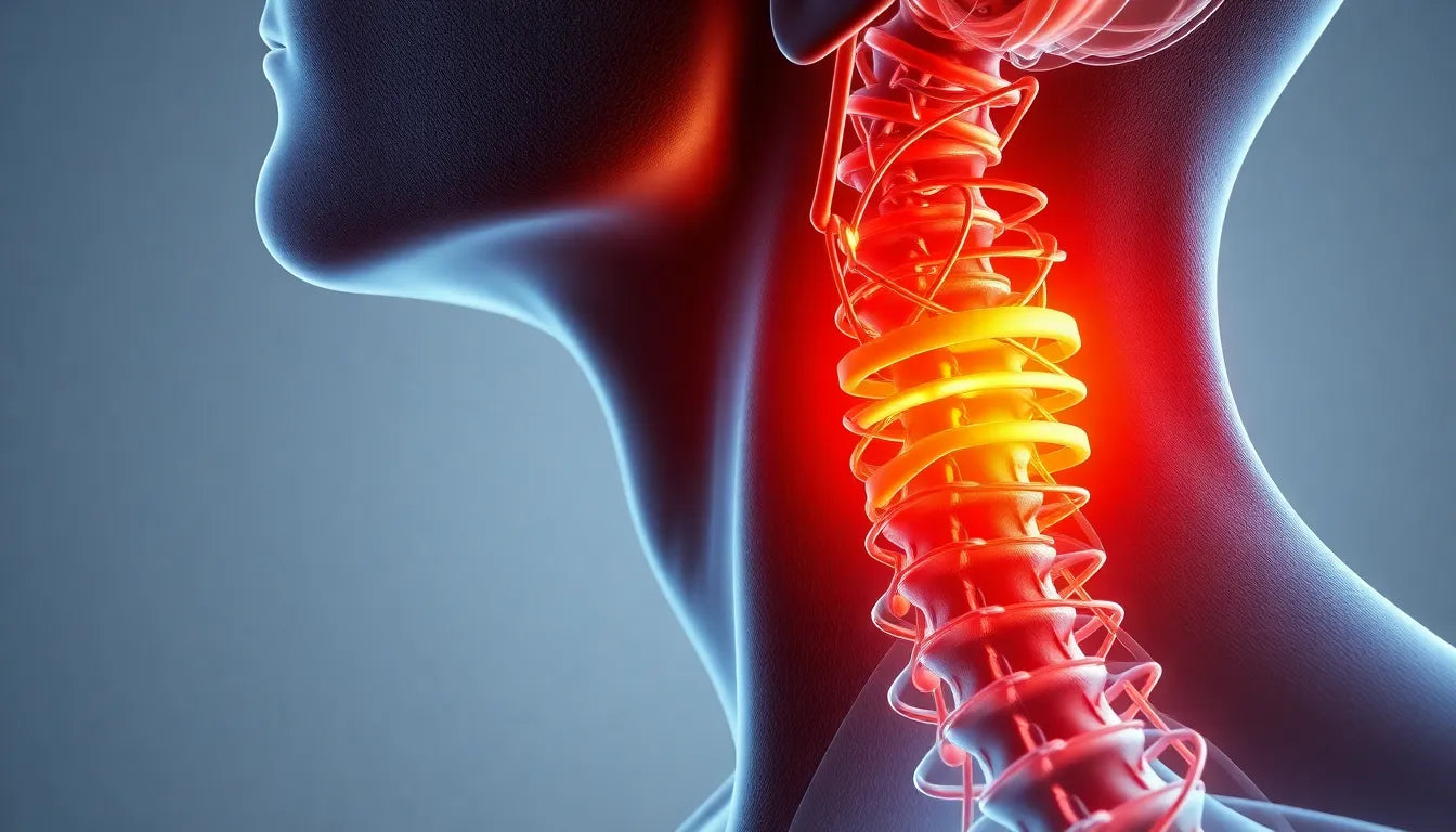
Back pain is a common ailment that affects millions of people worldwide, often disrupting daily activities and diminishing quality of life. For many, the root cause of this discomfort is a herniated disc, a condition that requires precise diagnosis to ensure effective treatment. The role of accurate diagnosis cannot be overstated, as it guides healthcare providers in crafting tailored treatment plans that address the specific needs of each patient.
Understanding MRI technology
Magnetic Resonance Imaging (MRI) is a powerful diagnostic tool that plays a crucial role in modern medicine. Unlike X-rays or CT scans, which rely on radiation, MRI uses magnetic fields and radio waves to produce detailed images of the body's internal structures. This non-invasive technique is particularly valuable for examining soft tissues, making it ideal for diagnosing spinal conditions such as herniated discs.
When it comes to spinal health, MRI scans offer unparalleled insights. They provide high-resolution images that allow medical professionals to assess the spine's intricate anatomy and pinpoint issues that might not be visible through other imaging methods. For individuals suffering from back pain, an MRI scan can reveal the presence, location, and severity of a herniated disc, offering a clear path forward for treatment.
The importance of accurate diagnosis
An accurate diagnosis is the cornerstone of effective treatment for herniated discs. Identifying the precise nature of the problem enables healthcare providers to recommend the most appropriate interventions, whether surgical or non-surgical. Conversely, a misdiagnosis or delayed diagnosis can lead to ineffective treatments, prolonged pain, and even potential complications.
In the case of herniated discs, timely and accurate identification of the issue can significantly impact a patient's recovery journey. It ensures that the treatment plan is specifically tailored to address the unique aspects of the condition, thereby enhancing the likelihood of successful outcomes and a return to normal activities.
In conclusion, MRI scans are invaluable in the diagnosis of herniated discs, offering a precise and detailed view of the spine's condition. By facilitating accurate diagnoses, MRI technology plays a crucial role in guiding effective treatment plans, ultimately improving the quality of life for those affected by back pain.
how mri technology diagnoses herniated discs
An MRI scan is a sophisticated process that provides a detailed view of the spine, making it an essential tool for diagnosing herniated discs. Unlike X-rays and CT scans, which are more effective for viewing bones, MRI excels at imaging soft tissues, including discs, nerves, and the spinal cord. This capability is crucial for identifying the presence and extent of a herniated disc, as well as its impact on surrounding nerves.
The MRI process involves the patient lying inside a large, tube-shaped magnet. The machine generates a powerful magnetic field and radio waves, which interact with the body's hydrogen atoms to produce detailed images. These images are then analyzed by radiologists to detect abnormalities in the spine. The spatial resolution of MRI is exceptional, often cited with a diagnostic accuracy rate of 95-97%, making it the gold standard for diagnosing spinal conditions like herniated discs.
advantages of mri over other imaging techniques
When it comes to diagnosing herniated discs, MRI offers several advantages over other imaging methods. While CT scans and X-rays are useful for assessing bone structures, they fall short in visualizing soft tissues. MRI, on the other hand, provides a comprehensive view of the spine's soft tissues, allowing for a more accurate diagnosis. This is particularly important for identifying the precise location, size, and severity of a herniated disc.
Moreover, MRI is invaluable for pre-operative planning. By providing detailed images of the spine, surgeons can plan interventions with greater precision, potentially leading to improved surgical outcomes and faster recovery times. MRI's ability to differentiate between various types of spinal conditions also aids in tailoring treatment plans to the specific needs of the patient, whether surgical or non-surgical.
real-world impact: a case study
Consider a hypothetical case of a patient named John, who has been experiencing chronic back pain and numbness in his leg. Despite trying various conservative treatments, his symptoms persist. An MRI scan reveals a herniated disc compressing a nerve root, pinpointing the exact cause of his discomfort. With this accurate diagnosis, John's healthcare team can develop a targeted treatment plan, potentially involving minimally invasive surgery to relieve the pressure on the nerve.
This scenario highlights how MRI can significantly alter the course of treatment. By providing a clear and detailed picture of the spinal condition, MRI enables healthcare providers to make informed decisions, ultimately improving patient outcomes and quality of life.
In summary, MRI scans play a pivotal role in diagnosing herniated discs, offering unmatched accuracy and detail. Their ability to visualize soft tissues and aid in treatment planning makes them indispensable in modern spinal healthcare. As technology continues to advance, MRI's capabilities are likely to expand, further enhancing its role in diagnosing and treating spinal conditions.
The role of MRI in treatment and recovery
Magnetic Resonance Imaging (MRI) is not only crucial for diagnosing herniated discs but also plays a significant role in planning effective treatment and facilitating recovery. The detailed images provided by MRI allow healthcare professionals to design precise surgical interventions, which can lead to improved surgical outcomes. By accurately pinpointing the affected areas and understanding the severity of the condition, surgeons can perform minimally invasive procedures that reduce recovery time and enhance patient comfort.
Moreover, MRI findings can guide non-surgical treatments, such as physical therapy or targeted injections, by identifying the exact location and nature of the disc herniation. This targeted approach ensures that treatments are more effective, reducing the likelihood of persistent symptoms or the need for further interventions.
Recent advancements in MRI technology
The field of MRI technology is continuously evolving, with recent advancements enhancing both diagnostic capabilities and patient experience. Innovations such as faster imaging techniques and higher-resolution scans allow for more detailed visualization of spinal structures, potentially leading to earlier and more accurate diagnoses. These advancements can help detect subtle changes in the spine that might have been overlooked with older technology.
In addition, developments in MRI machine design have improved patient comfort, reducing the noise and claustrophobia often associated with traditional MRI scans. Open MRI machines and shorter scan times are examples of innovations that make the procedure more accessible and less daunting for patients.
Looking to the future, continued advancements in MRI technology may include the integration of artificial intelligence to assist radiologists in interpreting scans more quickly and accurately. Such innovations could further enhance the role of MRI in diagnosing and treating spinal conditions, offering new hope for patients with herniated discs.
Frequently Asked Questions
What is a herniated disc, and what causes it?
A herniated disc occurs when the soft inner gel of a spinal disc protrudes through a tear in its outer layer. This condition can result from injury, heavy lifting, or age-related degeneration of the spinal discs. Common causes include repetitive strain, traumatic events, or simply the natural wear and tear of aging.
When should one consider getting an MRI for back pain?
An MRI is recommended for back pain when the pain is persistent, severe, or accompanied by neurological symptoms such as numbness or weakness. It is also advisable when conservative treatments, like rest or physical therapy, do not alleviate the symptoms, or when a more serious underlying condition is suspected.
Are there any risks associated with MRI scans?
MRI scans are generally safe and do not involve exposure to radiation, unlike X-rays or CT scans. However, individuals with certain implants, such as pacemakers, should inform their healthcare provider, as the strong magnetic fields can interfere with these devices.
How should I prepare for an MRI scan?
Before an MRI scan, patients should remove any metal objects, such as jewelry or watches, and wear comfortable clothing without metal fasteners. It's also important to inform the technician of any medical implants or conditions that might affect the scan. For those anxious about the procedure, relaxation techniques or mild sedatives can be discussed with the healthcare provider.
What can I expect during and after the MRI scan?
During the MRI scan, patients will lie still on a table that slides into the MRI machine. The scan typically lasts between 30 to 60 minutes, during which patients may hear loud tapping or thumping noises. Earplugs or headphones are usually provided to minimize discomfort. After the scan, patients can resume normal activities immediately, as there is no recovery time required.
```html
Sources
- Deuk Spine. (n.d.). "How to Read the MRI for a Herniated Disc | 5 Treatments."
- Ezra. (n.d.). "Understanding Back Pain and How an MRI of the Spine Can Help."
- Dr. Kevin Pauza. (n.d.). "What is a Herniated Disc MRI, and When Do You Need It?"
- Touchstone Imaging. (n.d.). "How Can a Lumbar Spine MRI Help to Diagnose Herniated Discs?"