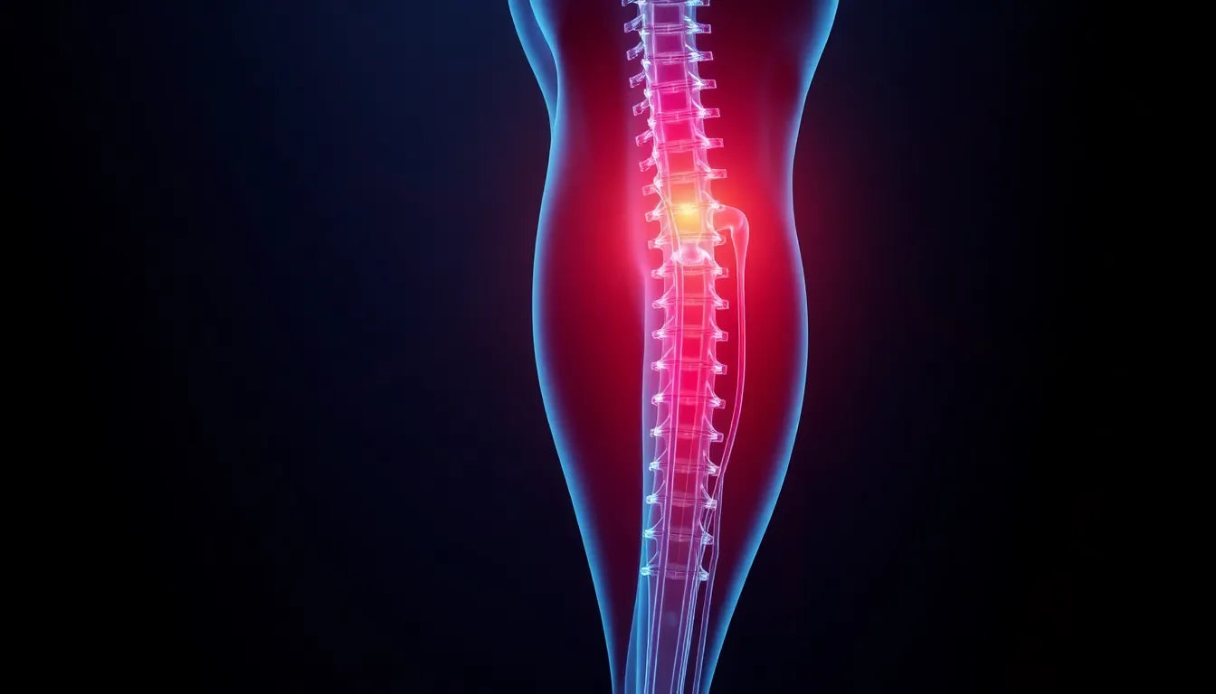
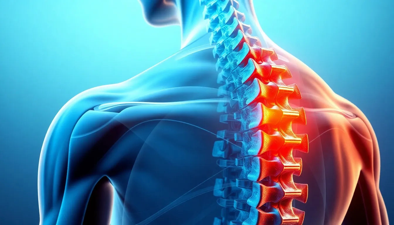
Understanding the complexities of spinal health is crucial, especially when dealing with issues like a herniated disc. A herniated disc, often referred to as a slipped or ruptured disc, occurs when the soft center of a spinal disc pushes through a crack in the tougher exterior casing. This condition can lead to discomfort, pain, and even nerve irritation, manifesting in symptoms such as back pain, numbness, or weakness in limbs. The causes of herniated discs can vary, ranging from age-related wear and tear to sudden injuries or strain.
Herniated discs are a prevalent concern, particularly among adults. Statistics reveal that approximately 1 to 2 percent of people will experience a herniated disc at some point in their lives, with the lumbar region being the most commonly affected area. Given its frequency and the potential for significant pain and discomfort, obtaining an accurate diagnosis is paramount. A precise diagnosis not only facilitates effective treatment but also plays a critical role in managing pain and preventing further complications.
Imaging techniques for herniated disc diagnosis
In the realm of medical diagnostics, imaging techniques are indispensable tools for identifying herniated discs. These methods allow healthcare professionals to visualize the spine's structure and pinpoint the exact location and extent of the disc herniation. Among the various imaging options available, Magnetic Resonance Imaging (MRI) stands out as the primary focus due to its unparalleled ability to provide detailed images of soft tissues, including spinal discs and nerves.
While MRI is the gold standard, other imaging techniques such as CT scans and X-rays also play a role in the diagnostic process. However, their effectiveness is generally limited compared to MRI, especially when it comes to evaluating soft tissues. CT scans and X-rays are often reserved for cases where MRI is not feasible or when additional information is needed to complement MRI findings.
The role of MRI in herniated disc diagnosis
So, why is MRI considered the gold standard for diagnosing herniated discs? Its reputation stems from its ability to produce high-resolution images that offer a comprehensive view of the spine's soft tissues. This capability is crucial for identifying the nuances of a herniated disc, such as its size, location, and impact on surrounding nerves. As you consider the importance of choosing the right diagnostic tool, imagine the peace of mind that comes with knowing your diagnosis is based on the most accurate and detailed imaging available. This assurance is why MRI remains the preferred choice for healthcare providers and patients alike.
MRI: the gold standard for herniated disc diagnosis
Magnetic Resonance Imaging (MRI) is widely recognized as the gold standard for diagnosing herniated discs, and for good reason. Its ability to provide high-resolution images of the spine’s soft tissues is unparalleled, allowing for detailed visualization of intervertebral discs and surrounding nerves. This precision is crucial for identifying not only the presence of a herniated disc but also its exact location, size, and the degree to which it may be impinging on nearby nerve structures.
One of the standout features of MRI is its accuracy. Studies have shown that MRI has a diagnostic accuracy rate of approximately 95% when it comes to identifying herniated discs. This high level of accuracy ensures that healthcare providers can make informed decisions regarding treatment plans, reducing the risk of misdiagnosis and unnecessary interventions. Furthermore, MRI's non-invasive nature makes it a safer option for patients, as it does not involve radiation exposure, unlike some other imaging techniques.
Alternative imaging techniques and their limitations
While MRI is the preferred method for diagnosing herniated discs, other imaging techniques like CT scans and X-rays are sometimes employed, albeit with certain limitations. CT scans, for instance, are useful in providing detailed images of bone structures and can be helpful in cases where MRI is contraindicated, such as in patients with certain metal implants. However, CT scans are less effective than MRI when it comes to evaluating soft tissues, which are crucial in diagnosing herniated discs.
X-rays, on the other hand, are limited in their ability to visualize soft tissues and are typically used to rule out other conditions that may cause similar symptoms, such as fractures or tumors. In some cases, these alternative imaging methods may be used in conjunction with MRI to provide a more comprehensive assessment of the spine.
The clinical process: combining imaging with patient history
The process of diagnosing a herniated disc is not solely reliant on imaging techniques. A thorough clinical evaluation, including a detailed patient history and physical examination, is essential. Healthcare providers will assess the patient's symptoms, such as the location and severity of pain, and any neurological deficits. This information, combined with imaging results, provides a holistic view of the patient's condition.
In cases where surgical intervention is considered, MRI plays a pivotal role in surgical planning. The detailed images provided by MRI help surgeons understand the anatomy of the affected area, allowing for precise surgical approaches that minimize risks and improve outcomes.
Comparative analysis of imaging techniques
To better understand the strengths and weaknesses of various imaging techniques, consider the following comparison:
| Imaging Type | Detail Level | Invasiveness | Accuracy | Cost |
|---|---|---|---|---|
| MRI | High | Non-invasive | 95% | Moderate to High |
| CT Scan | Moderate | Non-invasive | Less than MRI | Moderate |
| X-ray | Low | Non-invasive | Low for soft tissues | Low |
This table underscores MRI's superiority in detail and accuracy, particularly for soft tissue evaluation, which is critical in diagnosing herniated discs. While CT scans and X-rays have their place in the diagnostic process, their limitations in soft tissue visualization make MRI the preferred choice for most cases.
In conclusion, the power of MRI in diagnosing herniated discs cannot be overstated. Its ability to provide detailed and accurate images of the spine's soft tissues makes it an indispensable tool in the diagnostic arsenal, ensuring patients receive the most effective and targeted treatment plans.
The role of MRI in treatment and recovery
Once a herniated disc is accurately diagnosed through MRI, the findings significantly influence the treatment plan. MRI results provide a detailed map of the affected area, allowing healthcare providers to tailor treatment options that best suit the patient's condition. Non-surgical treatments, such as physical therapy, pain management, and lifestyle modifications, are often the first line of defense. These approaches aim to alleviate symptoms and improve mobility without the need for invasive procedures.
In addition to guiding treatment, MRI findings play a crucial role in recovery. For instance, understanding the extent of the herniation and its impact on surrounding tissues helps in devising effective rehabilitation strategies. This can include targeted physical therapy exercises aimed at strengthening the back muscles and improving flexibility, which are essential for preventing future injuries.
Ergonomic solutions for support and recovery
Ergonomic solutions are vital in supporting recovery and managing pain post-diagnosis. Products such as ergonomic chairs, lumbar support cushions, and adjustable desks can help maintain proper posture, reducing strain on the spine. These aids are particularly beneficial for individuals who spend long hours sitting or standing, as they promote spinal alignment and alleviate pressure on the herniated disc.
Incorporating ergonomic solutions into daily routines not only supports recovery but also helps prevent further disc-related issues. By minimizing stress on the spine and encouraging healthy movement patterns, these tools serve as an integral part of a comprehensive treatment plan for herniated disc patients.
Frequently asked questions
What does a herniated disc look like on an MRI?
A herniated disc on an MRI typically appears as a bulge or protrusion in the spinal disc, often compressing nearby nerves. Radiologists look for these bulges and any displacement of the disc material, which can indicate the severity of the herniation and its potential impact on surrounding structures.
Is MRI the best scan for herniated discs?
MRI is widely regarded as the best scan for herniated discs due to its ability to provide detailed images of soft tissues, including discs and nerves. While other scans like CT or X-rays can be useful in specific situations, MRI remains the preferred choice for its accuracy and non-invasive nature.
Can CT scans diagnose herniated discs?
CT scans can diagnose herniated discs, but they are generally less effective than MRIs for evaluating soft tissues. CT scans are more often used when MRI is not available or contraindicated, such as in patients with certain metal implants.
How long does an MRI for a herniated disc take?
An MRI for a herniated disc typically takes about 30 to 60 minutes, including preparation and scanning time. The exact duration may vary depending on the specific protocol used and the area being examined.
Are there any risks associated with MRI scans?
MRI scans are generally considered safe and do not involve radiation exposure. However, they may not be suitable for patients with certain implants, such as pacemakers or metal fragments. It's important for patients to inform their healthcare provider of any implants or medical conditions before undergoing an MRI.
In summary, the use of MRI in diagnosing and managing herniated discs is invaluable. Its detailed imaging capabilities ensure precise diagnoses, guiding effective treatment and recovery plans. By combining MRI findings with ergonomic solutions, patients can achieve optimal outcomes and maintain spinal health.
Sources
- Deuk Spine. (n.d.). "MRI for Herniated Discs."
- Spine-health. (n.d.). "MRI Scan for Lumbar Herniated Disc."
- University of Maryland Medical Center. (n.d.). "Herniated Disc."
- NYU Langone Health. (n.d.). "Herniated Disc in Adults: Diagnosis."
- AMA Journal of Ethics. (2013). "The Ethics of Imaging for Spinal Conditions."