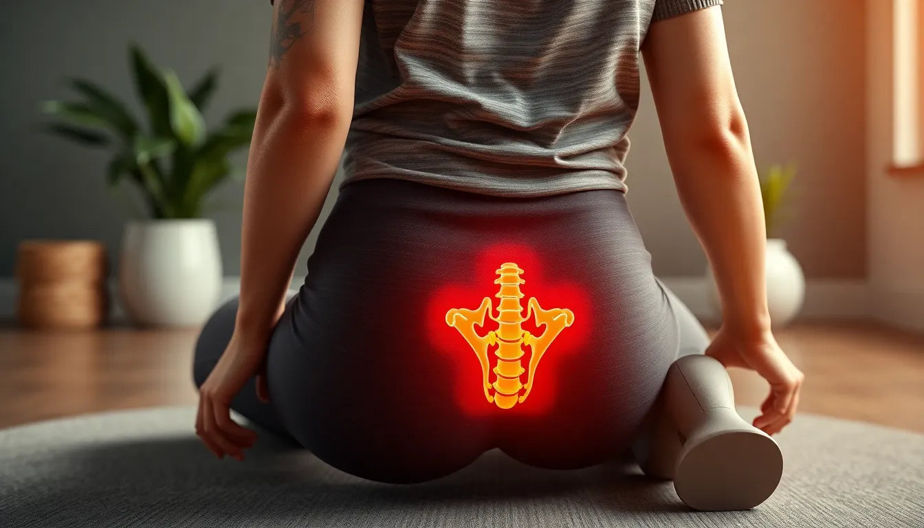
Back pain is a common ailment that affects millions of people worldwide, often disrupting day-to-day activities and diminishing quality of life. Among the various causes of back pain, a herniated disc stands out due to its prevalence and the significant discomfort it can cause. A herniated disc occurs when the soft inner gel of a spinal disc bulges out through a tear in the tougher outer layer. This condition can lead to symptoms such as sharp pain, numbness, and weakness, typically affecting the back and extending to the arms or legs depending on the location of the herniation.
Understanding the complexity of back pain
The complexity of back pain lies in its myriad causes and manifestations, making it crucial to accurately diagnose the underlying issue. A herniated disc is often suspected when patients present with specific symptoms, but its diagnosis can be challenging due to overlapping symptoms with other spinal conditions, such as spinal stenosis or degenerative disc disease. This complexity underscores the importance of a precise diagnosis to ensure effective treatment and management of the condition, thereby alleviating pain and restoring mobility.
The quest for accurate diagnosis
Diagnosing a herniated disc requires a methodical approach, as the symptoms can mimic those of other conditions. The quest for an accurate diagnosis is not only about identifying the presence of a herniation but also understanding its impact on surrounding nerves and tissues. This blog post aims to demystify the diagnostic process for a herniated disc, providing insight into the steps healthcare professionals take to differentiate it from other spinal disorders. By understanding these diagnostic procedures, individuals can gain clarity on their condition and the path to recovery.
Initial diagnostic steps for a herniated disc
When a patient presents with symptoms indicative of a herniated disc, the diagnostic journey begins with a thorough medical history and physical examination. Understanding the patient's symptoms, lifestyle, and medical background is crucial. Physicians typically ask about the onset and duration of symptoms, any activities that exacerbate or alleviate pain, and previous medical conditions that might contribute to spinal issues. This information provides a foundation for identifying the root cause of the discomfort and guides further diagnostic steps.
The physical examination involves several tests to assess nerve function and pinpoint areas of discomfort. One common test is the straight leg raise test, which helps determine if there is nerve compression in the lower back. During this test, the patient lies flat while the doctor gently lifts each leg. Pain or discomfort during this maneuver can indicate nerve irritation, often associated with a herniated disc. Additional assessments may include checking reflexes, muscle strength, and sensation to identify any areas of weakness or numbness.
Advanced imaging tests to confirm diagnosis
Once the initial examination suggests a herniated disc, advanced imaging tests are employed to confirm the diagnosis and determine the extent of the condition. Magnetic Resonance Imaging (MRI) is the gold standard for diagnosing herniated discs, as it provides detailed images of the spinal anatomy, including the discs, nerves, and surrounding soft tissues. MRI scans can reveal the exact location and severity of the herniation, offering invaluable information for treatment planning.
For patients who cannot undergo an MRI due to contraindications such as metal implants or severe claustrophobia, a Computed Tomography (CT) scan serves as an alternative. CT scans provide a three-dimensional view of the spine, allowing doctors to visualize bone structures and detect any abnormalities. Although CT scans are less detailed than MRIs in showing soft tissues, they are instrumental in diagnosing herniated discs in certain cases.
X-rays, while not capable of visualizing soft tissues like discs, play a role in ruling out other bone-related issues such as fractures or spondylolisthesis that might contribute to back pain. They are often used as an initial imaging test to assess the overall alignment and integrity of the spinal column.
Another specialized imaging test, the myelogram, involves injecting a contrast dye into the spinal fluid to enhance the visibility of the spinal cord and nerves on X-rays or CT scans. This test is particularly useful in complex cases where other imaging tests do not provide sufficient information.
Comprehensive approach to diagnosing herniated discs
Accurate diagnosis of a herniated disc relies on a comprehensive approach that combines medical history, physical examination, and advanced imaging tests. This multifaceted strategy ensures that the diagnosis is precise and tailored to the individual's specific condition. Healthcare professionals play a crucial role in interpreting the results of these tests, guiding patients through the diagnostic process, and developing a personalized treatment plan.
Incorporating visual aids, such as diagrams of the spine and images of diagnostic tests, can enhance patient understanding of their condition. Additionally, comparing the different diagnostic tests in a table format can help highlight their specific uses, benefits, and limitations, providing a clearer picture of the diagnostic process.
Understanding the diagnostic steps for a herniated disc empowers patients to actively participate in their healthcare journey. By being informed about the process, individuals can engage in meaningful discussions with their healthcare providers, leading to better outcomes and a clearer path to recovery.
Additional diagnostic tests for a herniated disc
Beyond the initial and imaging tests, other diagnostic methods are employed to provide a comprehensive understanding of a herniated disc. Electromyogram (EMG) and Nerve Conduction Studies (NCS) are instrumental in measuring the electrical activity of nerves and muscles. These tests can identify nerve damage or compression, which is crucial for pinpointing the precise location and extent of nerve involvement.
In certain cases, laboratory tests such as Erythrocyte Sedimentation Rate (ESR) and C-reactive Protein (CRP) are conducted to rule out other conditions like infections or inflammatory diseases that could mimic the symptoms of a herniated disc. These tests help in differentiating between a herniated disc and other potential causes of back pain.
Comprehensive diagnostic approach
An accurate diagnosis of a herniated disc requires a holistic approach that integrates medical history, physical examination, advanced imaging, and additional diagnostic tests. This comprehensive strategy ensures that the diagnosis is both precise and personalized. Healthcare professionals are pivotal in interpreting these test results, forming a clear diagnosis, and crafting a tailored treatment plan to address the patient's specific needs.
Visual aids, such as diagrams of the spine and images of diagnostic tests, can significantly enhance a patient's understanding of their condition. A comparison table of different diagnostic tests can further clarify their specific uses, benefits, and limitations, providing a clearer picture of the diagnostic process.
Frequently Asked Questions
What symptoms indicate a herniated disc?
Common symptoms of a herniated disc include back pain, leg pain, numbness, tingling, and weakness, particularly in the arms or legs. These symptoms can vary depending on the location of the herniation and the nerves affected.
How long does the diagnostic process take?
The diagnostic process for a herniated disc can vary in duration. Typically, it involves an initial consultation, followed by physical examinations and imaging tests. The entire process can take anywhere from a few days to a couple of weeks, depending on the availability of tests and specialists.
Are the diagnostic tests painful?
Most diagnostic tests for a herniated disc, such as MRI and CT scans, are non-invasive and painless. However, tests like the EMG may cause some discomfort due to needle insertion, but it is generally well-tolerated.
What should I expect during an MRI scan?
During an MRI scan, you will lie still on a table that slides into a large, tube-like machine. The procedure is painless and typically lasts 30 to 60 minutes. It is important to remain still to ensure clear images. If you are claustrophobic, inform your doctor, as they may provide a sedative to help you relax.
Can a herniated disc heal without surgery?
Yes, many herniated discs can heal without the need for surgery. Conservative treatments, including physical therapy, medications, and lifestyle modifications, are often effective. The treatment plan is personalized based on the diagnosis and the severity of symptoms.