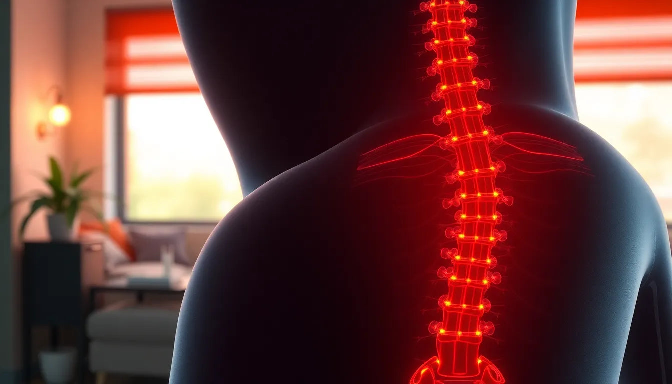
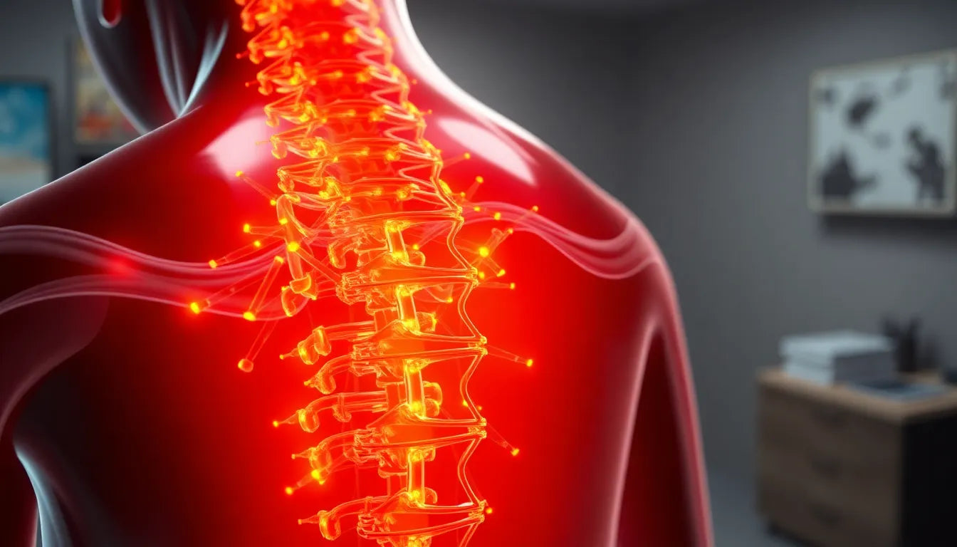
Understanding the intricacies of a herniated disc diagnosis begins with grasping the basics of what a herniated disc actually is. This condition occurs when the soft inner gel of a spinal disc pushes through a crack in the tougher exterior casing. The result can be irritation or compression of nearby nerves, leading to symptoms such as back pain, numbness, or weakness in the limbs. These symptoms can vary widely in intensity and can significantly impact daily life, making an accurate diagnosis crucial for effective treatment.
Why accurate diagnosis matters
Diagnosing a herniated disc can be a complex process due to the overlapping symptoms it shares with other spinal conditions. The pain and discomfort associated with a herniated disc might be mistaken for other back issues, such as a muscle strain or arthritis. This complexity underscores the importance of a precise and comprehensive diagnostic approach. Without an accurate diagnosis, treatment plans may not address the root cause of the symptoms, potentially leading to prolonged discomfort or ineffective treatment outcomes.
The purpose of this blog post is to delve into the diagnostic processes that healthcare professionals employ to identify a herniated disc with precision. By understanding these processes, patients can gain insight into what to expect during their medical evaluations and the importance of each diagnostic step. From initial assessments to advanced imaging techniques, each step plays a vital role in painting a comprehensive picture of the spinal health of a patient.
In the upcoming sections, we will explore the initial diagnostic steps, including the significance of medical history and physical examinations, and delve into advanced imaging techniques like MRI and CT scans. These methods are pivotal in confirming a herniated disc diagnosis and guiding the subsequent treatment plan. By shedding light on these diagnostic processes, we aim to empower readers with the knowledge needed to navigate their own healthcare journeys effectively.
Initial diagnostic steps: medical history and physical examination
The journey to accurately diagnosing a herniated disc often begins with a thorough review of the patient's medical history and a detailed physical examination. Understanding the patient's history is crucial as it provides context to the symptoms presented. Patients are typically asked about the onset, duration, and nature of their symptoms, as well as any previous injuries or medical conditions that could contribute to their current state.
During the physical examination, healthcare providers perform specific tests to evaluate nerve function and pain response. These tests might include checking reflexes, muscle strength, and sensory responses in the affected areas. The doctor may also ask the patient to perform certain movements to identify any limitations or pain triggers. This initial assessment helps to rule out other potential causes of back pain and narrows down the possibility of a herniated disc.
Advanced imaging techniques: mri and ct scans
When initial assessments suggest the presence of a herniated disc, advanced imaging techniques become essential in confirming the diagnosis. Magnetic Resonance Imaging (MRI) is often considered the gold standard for diagnosing herniated discs due to its ability to produce detailed images of soft tissues, including the spinal discs and nerves. An MRI can reveal the exact location and extent of the herniation, providing critical information for developing an effective treatment plan.
In some cases, a Computed Tomography (CT) scan may be used, especially when MRI is not available or suitable for the patient. CT scans provide a detailed view of the spine's bone structures and can be helpful in identifying any associated bone abnormalities. While CT scans are less detailed than MRIs in terms of soft tissue visualization, they play a valuable role in certain diagnostic scenarios, particularly when assessing the relationship between the herniated disc and the surrounding bony structures.
Other imaging options: x-rays and myelograms
Although X-rays are not typically used to diagnose herniated discs directly, they can help rule out other conditions, such as fractures or tumors, that might cause similar symptoms. X-rays provide a clear view of the spine's alignment and can detect any significant structural changes.
Myelograms, on the other hand, involve injecting a contrast dye into the spinal canal before taking X-ray or CT images. This technique can highlight abnormalities in the spinal cord or nerve roots, making it a useful tool when MRI is contraindicated or when additional detail is needed to assess the extent of nerve compression.
Nerve function tests: emg and ncs
To complement imaging tests, nerve function tests such as Electromyography (EMG) and Nerve Conduction Studies (NCS) are often performed. These tests assess the electrical activity of muscles and the speed of nerve signal transmission, respectively. EMG and NCS can help determine the extent of nerve damage or compression caused by a herniated disc, providing a comprehensive view of the condition's impact on nerve function.
By combining these diagnostic tools, healthcare professionals can achieve a precise and thorough understanding of the patient's condition, ensuring that the chosen treatment plan addresses all aspects of the herniated disc. This comprehensive approach not only aids in confirming the diagnosis but also plays a pivotal role in guiding effective treatment strategies.
Comparative analysis of diagnostic tests
When diagnosing a herniated disc, healthcare professionals employ various diagnostic tests, each with its own strengths and limitations. Below is a comparative analysis of the most commonly used diagnostic tools:
| Diagnostic Test | Pros | Cons |
|---|---|---|
| MRI (Magnetic Resonance Imaging) | Provides detailed images of soft tissues; non-invasive; no radiation exposure. | Expensive; not suitable for patients with certain implants. |
| CT Scan (Computed Tomography) | Excellent for visualizing bone structures; quicker than MRI. | Less detail on soft tissues compared to MRI; involves radiation exposure. |
| X-ray | Good for identifying bone abnormalities; widely available and cost-effective. | Limited in diagnosing soft tissue issues like herniated discs. |
| Myelogram | Useful when MRI is contraindicated; highlights nerve root and spinal cord issues. | Invasive; involves radiation and potential allergic reactions to contrast dye. |
The choice of diagnostic test depends on individual patient circumstances, including medical history, symptoms, and any contraindications for specific procedures. For instance, while MRI is often preferred for its detailed soft tissue images, a CT scan might be more appropriate for patients with metal implants.
Patient experience and testimonials
For many patients, the journey to diagnosing a herniated disc can be daunting. However, accurate diagnosis can significantly impact treatment outcomes. Jane, a 45-year-old patient who underwent an MRI, shared, "The MRI was crucial in pinpointing the exact location of my herniated disc. It gave my doctor the information needed to tailor my treatment plan, which made a huge difference in my recovery."
Similarly, Tom, who had a CT scan, noted, "I was initially apprehensive about the procedure, but it was quick and provided clear results that helped my doctor rule out other conditions."
These testimonials highlight the importance of choosing the right diagnostic approach, as it can lead to more effective and personalized treatment strategies.
Visual aids for understanding
Incorporating visual aids, such as diagrams of the spine and images from diagnostic tests, can enhance understanding of herniated discs and the diagnostic process. Visual representations can clarify how herniated discs affect spinal structures and nerves, making it easier for patients to comprehend their condition and the necessity of certain tests.
Conclusion
Diagnosing a herniated disc with precision involves a multifaceted approach that combines patient history, physical examinations, and advanced diagnostic tests. Each method, from MRI to nerve function tests, plays a crucial role in confirming the diagnosis and guiding effective treatment plans. Understanding these processes empowers patients to make informed decisions about their healthcare journey. By choosing the appropriate diagnostic approach, patients and healthcare providers can work together to achieve optimal treatment outcomes.
Frequently Asked Questions
What are the symptoms of a herniated disc?
Common symptoms include back pain, numbness, tingling, and weakness in the limbs. These symptoms can vary in intensity and location depending on the affected nerves.
How long does it take to diagnose a herniated disc?
The timeline for diagnosis can vary, but typically it involves an initial consultation, followed by physical examinations and imaging tests. This process can take anywhere from a few days to a couple of weeks, depending on the availability of diagnostic services.
Is MRI the only way to diagnose a herniated disc?
While MRI is often preferred for its detailed images of soft tissues, other diagnostic options include CT scans, X-rays, and myelograms. The choice depends on the patient's specific circumstances and medical history.
Can a herniated disc heal without surgery?
Yes, many herniated discs can heal with non-surgical treatments such as physical therapy, medication, and lifestyle modifications. Surgery is typically considered when conservative treatments fail to alleviate symptoms.
What should I expect during a diagnostic test for a herniated disc?
During imaging tests like MRI or CT scans, you will be asked to lie still while the machine captures detailed images of your spine. Nerve function tests, such as EMG and NCS, involve minor electrical impulses to assess nerve and muscle function.