


When it comes to diagnosing spinal issues, many people believe that an X-ray is the go-to solution. However, this common misconception can lead to misunderstandings, particularly when dealing with complex conditions like a herniated disc. While X-rays are a valuable tool in the medical field, they are not always the definitive answer for every spinal problem. Understanding the limitations of X-rays and the importance of accurate diagnosis is crucial for effective treatment.
Understanding herniated discs
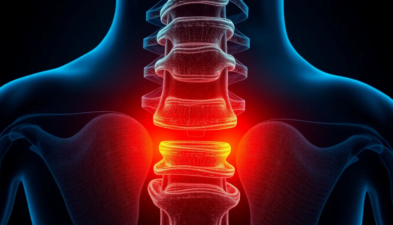
A herniated disc, also known as a slipped or ruptured disc, occurs when the soft inner gel of a spinal disc pushes through a tear in its tougher exterior. This condition can lead to a range of symptoms, including pain, numbness, and tingling, often radiating down the arms or legs depending on the location of the herniation. Herniated discs are relatively common, especially in individuals between the ages of 30 and 50, and can significantly impact daily life, making even simple tasks challenging.
The prevalence of herniated discs highlights the need for precise diagnosis. Misdiagnosis or delayed diagnosis can lead to prolonged discomfort and ineffective treatment plans. Therefore, it's essential to utilize the right diagnostic tools to confirm the presence and severity of a herniated disc.
The purpose of this post
In this post, we aim to explore whether X-rays can effectively diagnose a herniated disc and discuss alternative diagnostic methods that provide more accurate results. By delving into the capabilities and limitations of X-rays, as well as other imaging techniques, we hope to offer a clearer understanding of how to approach the diagnosis of herniated discs. This knowledge is vital for anyone experiencing symptoms or seeking to understand more about spinal health and the best practices for diagnosis and treatment.
Diagnostic limitations of X-rays in identifying herniated discs
When it comes to diagnosing a herniated disc, X-rays fall short due to their inherent limitations. X-rays are primarily designed to capture images of dense structures like bones, which means they do not effectively visualize soft tissues such as spinal discs and nerves. This limitation is crucial because herniated discs involve the displacement of soft tissue, which X-rays cannot adequately capture.
Medical institutions like NYU Langone Health and the Hospital for Special Surgery (HSS) emphasize that while X-rays can rule out other potential causes of pain, such as fractures or tumors, they are not suitable for diagnosing herniated discs. X-rays are valuable for assessing the alignment of the spine and identifying structural abnormalities, but they cannot provide the detailed images necessary to confirm a herniated disc. Understanding these limitations is essential for patients and healthcare providers to avoid misdiagnosis and ensure that the most effective diagnostic tools are used.
Exploring alternative diagnostic tests
To accurately diagnose a herniated disc, medical professionals often turn to advanced imaging techniques that provide detailed views of soft tissues. Among these, MRI scans are considered the gold standard. MRI, or Magnetic Resonance Imaging, offers comprehensive images of the spine, including the discs, nerves, and surrounding soft tissues. This level of detail is crucial for confirming the presence and extent of a herniated disc, making MRI scans highly effective for diagnosis.
In addition to MRI, CT scans and myelograms are also used in certain cases. CT scans provide cross-sectional images of the spine, offering a different perspective that can be useful in complex cases. Myelograms, which involve the injection of contrast material into the spinal canal, enhance the visibility of the spinal cord and nerve roots, providing additional detail that can aid in diagnosis. These alternative tests are often employed when MRI scans are not feasible or when additional information is needed to plan treatment.
Comparison table: diagnostic tools for herniated discs
| Diagnostic Tool | Effectiveness | Cost | Usage |
|---|---|---|---|
| X-ray | Low for soft tissues | Low | Rules out bone issues |
| MRI | High | High | Confirms herniated discs |
| CT Scan | Moderate | Moderate | Cross-sectional imaging |
| Myelogram | High with contrast | Moderate to high | Detailed nerve imaging |
Procedures and precautions for X-ray imaging
While X-rays are not ideal for diagnosing herniated discs, they remain a common diagnostic tool for other spinal issues. During an X-ray procedure, patients are typically positioned in a way that allows for clear imaging of the spine. It is important to remove any metal objects, such as jewelry or belts, that could interfere with the image quality.
Precautions are also important, especially for certain populations. For instance, pregnant women are generally advised to avoid X-rays unless absolutely necessary, due to the potential risks of radiation exposure to the developing fetus. Additionally, contrast materials used in some imaging tests, like myelograms, require careful consideration and preparation, including informing the technician of any allergies or medical conditions.
By understanding the limitations and appropriate applications of each diagnostic tool, patients and healthcare providers can work together to ensure accurate diagnosis and effective treatment planning for herniated discs and other spinal conditions.
The importance of accurate diagnosis for herniated discs
Accurate diagnosis is crucial when dealing with herniated discs, as it directly impacts the effectiveness of treatment plans. Using the right diagnostic tools ensures that the condition is correctly identified and appropriately managed. Misdiagnosis or delayed diagnosis can lead to prolonged discomfort, unnecessary treatments, and potentially worsening symptoms. By employing advanced imaging techniques such as MRI or CT scans, healthcare providers can gain a comprehensive understanding of the spinal condition, allowing for tailored treatment strategies that address the root cause of the symptoms.
Furthermore, accurate diagnosis helps in distinguishing herniated discs from other conditions with similar symptoms, such as spinal stenosis or degenerative disc disease. This differentiation is essential for developing a targeted treatment approach, whether it involves physical therapy, medication, or, in some cases, surgical intervention. Patients who receive a precise diagnosis can embark on a more effective recovery journey, improving their quality of life and reducing the risk of future complications.
Frequently Asked Questions
Can an X-ray detect a herniated disc?
No, an X-ray cannot detect a herniated disc. X-rays are designed to image dense structures like bones and cannot visualize soft tissues such as spinal discs and nerves. They are primarily used to rule out other conditions like fractures or tumors.
What are the best tests for diagnosing a herniated disc?
MRI scans are considered the gold standard for diagnosing herniated discs, as they provide detailed images of soft tissues, including discs and nerves. CT scans and myelograms can also be used in certain cases to provide additional information.
Why might a doctor order an X-ray if it can't show a herniated disc?
Doctors may order X-rays to rule out other potential causes of back pain, such as fractures, tumors, or structural abnormalities. X-rays help assess the alignment of the spine and ensure that no bone-related issues are contributing to the symptoms.
Are there any risks associated with MRI or CT scans?
MRI scans are generally safe and do not involve radiation. However, individuals with metal implants or certain medical devices may not be suitable candidates for MRI. CT scans involve exposure to a small amount of radiation, which is generally considered safe, but repeated exposure should be minimized. It's important to discuss any concerns with your healthcare provider.
How should I prepare for an MRI or CT scan?
Before an MRI or CT scan, you should remove any metal objects, such as jewelry or belts, as they can interfere with the imaging. Inform the technician of any medical conditions, allergies, or if you are pregnant. For some CT scans, you may need to fast for a few hours beforehand, especially if a contrast dye is used.
Conclusion
In conclusion, while X-rays are a valuable tool in diagnosing various spinal issues, they are not suitable for identifying herniated discs. Advanced imaging techniques like MRI and CT scans provide the necessary detail to accurately diagnose and plan treatment for herniated discs. It is essential to consult with healthcare professionals to ensure the correct diagnostic approach is taken, leading to effective treatment and improved patient outcomes.