Imagine waking up one morning with a sharp pain shooting down your leg. You wonder if this could be a sign of a herniated disc, a condition notorious for its ability to cause significant discomfort and disrupt daily life. Back pain is incredibly common, affecting millions of people worldwide, and understanding its root cause is crucial in managing symptoms effectively.
understanding a herniated disc
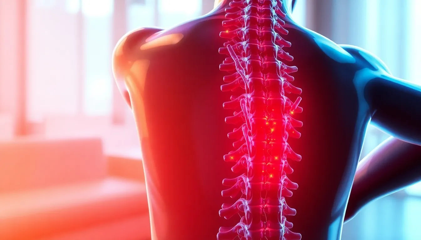
A herniated disc, sometimes referred to as a slipped or ruptured disc, occurs when the soft inner gel of a spinal disc pushes through a crack in the tougher exterior. This can irritate nearby nerves and result in pain, numbness, or weakness in an arm or leg. While not everyone with a herniated disc experiences symptoms, for those who do, the impact on daily activities can be profound. Accurate diagnosis is essential to tailor a treatment plan that alleviates pain and restores function.
the importance of accurate diagnosis
Diagnosing a herniated disc accurately is the first step toward effective management and relief. Without a proper diagnosis, treatment may be ineffective or even exacerbate the problem. The diagnostic process involves a combination of a detailed medical history, physical examination, and, when necessary, imaging tests. These steps help healthcare providers understand the extent of the issue and determine the best course of action.
The goal of this post is to unravel the diagnostic process for a herniated disc, providing clarity on what patients can expect when seeking medical attention for back pain. By understanding the steps involved in diagnosis, patients can be better prepared to discuss their symptoms with healthcare providers and make informed decisions about their treatment options.
diagnosis basics: the first steps
The journey to diagnosing a herniated disc begins with a thorough review of the patient's medical history and a comprehensive physical examination. This initial step is crucial, as it allows healthcare providers to gather essential information about the symptoms and their impact on daily life. Understanding the patient's history of back pain, previous injuries, and lifestyle factors can provide valuable context for the current condition.
During the physical examination, the healthcare provider will assess the patient's range of motion, reflexes, and muscle strength. They will also inquire about the nature of the pain—whether it's sharp, dull, or radiating—and any accompanying symptoms like numbness or tingling. This detailed evaluation helps in identifying potential nerve involvement and guides the subsequent steps in the diagnostic process.
physical examination techniques: straight leg raise test
One of the key physical examination techniques used to diagnose a herniated disc is the straight leg raise test. This simple yet effective test involves the patient lying flat on their back while the healthcare provider gently lifts one leg. If lifting the leg causes pain to shoot down the back of the leg, it may indicate nerve impingement associated with a herniated disc.
The significance of this test lies in its ability to pinpoint nerve root irritation, a common consequence of disc herniation. Although the straight leg raise test alone cannot confirm a herniated disc, it plays a pivotal role in the overall assessment, guiding the need for further diagnostic testing.
imaging techniques: mri, ct scans, and x-rays
When physical examination results suggest a herniated disc, imaging techniques become essential in confirming the diagnosis. Magnetic Resonance Imaging (MRI) is often the gold standard in visualizing disc herniation. It provides detailed images of the spinal discs, nerves, and surrounding soft tissues, making it particularly beneficial in complex cases where precise visualization is required.
In certain situations, a Computed Tomography (CT) scan may be employed, especially when MRI is contraindicated due to medical reasons. CT scans are effective in evaluating the bony structures of the spine and can offer vital insights when assessing calcified discs or spinal alignment issues.
Although X-rays are not capable of directly visualizing herniated discs, they are useful in evaluating spinal alignment and ruling out other potential causes of back pain, such as fractures or tumors. X-rays are often a preliminary step before more advanced imaging is pursued.
nerve function tests: emg and ncs
When symptoms suggest significant nerve damage, nerve function tests such as Electromyography (EMG) and Nerve Conduction Studies (NCS) may be conducted. These tests assess the electrical activity of muscles and the speed of nerve signal transmission, respectively. They are particularly useful in cases where the degree of nerve involvement needs to be quantified, aiding in the formulation of a comprehensive treatment plan.
advanced testing: myelogram
In rare and complex cases where standard imaging techniques do not provide conclusive results, a myelogram may be performed. This specialized test involves the injection of a contrast dye into the spinal canal, followed by X-ray or CT imaging. A myelogram is reserved for situations where detailed visualization of the spinal cord and nerve roots is necessary, often when surgical intervention is being considered.
clinical guidelines for imaging
It is important to note that not all back pain warrants immediate imaging. Clinical guidelines suggest that imaging should be reserved for cases with severe symptoms or red flags indicating neurological compromise, such as significant weakness or loss of bowel or bladder control. By adhering to these guidelines, unnecessary exposure to radiation and healthcare costs can be minimized, ensuring that imaging is used judiciously and effectively.
decision-making in diagnosis
Determining the appropriate diagnostic approach for a herniated disc involves a careful balance between clinical judgment and patient-specific factors. A decision tree can be a valuable tool in guiding healthcare providers through this process. Initially, the focus is on correlating the patient's symptoms with findings from the physical examination. If the symptoms align with potential nerve impingement, further testing may be warranted.
In cases where the patient's symptoms are mild and manageable, conservative management such as physical therapy may be recommended before pursuing advanced imaging. However, if the patient exhibits severe symptoms, such as significant weakness or loss of sensation, immediate imaging and specialist referral are crucial. By utilizing a structured approach, healthcare providers can ensure that each diagnostic step is necessary and beneficial.
patient education and managing expectations
Educating patients about the diagnostic process and the rationale behind each test is essential in managing expectations and reducing anxiety. It is important to explain why certain tests, like X-rays, may be limited in their ability to directly diagnose a herniated disc. Over-imaging can lead to unnecessary stress and healthcare costs, so emphasizing the importance of symptom correlation can help patients understand the clinical decision-making process.
While awaiting a definitive diagnosis, patients should be reassured that there are effective strategies to manage symptoms. Pain relief measures, physical therapy, and lifestyle modifications can significantly improve quality of life. By empowering patients with knowledge and practical advice, they can actively participate in their care and make informed decisions alongside their healthcare providers.
frequently asked questions
What if my MRI is normal but I still have pain?
It's possible for an MRI to appear normal even when pain persists. This can occur if the pain is due to muscle strain, ligament issues, or other non-disc-related causes. If pain continues despite normal imaging, seeking a second opinion or exploring other potential sources of discomfort with your healthcare provider is advisable.
When should I worry about leg numbness or weakness?
If you experience leg numbness or weakness, particularly if it worsens over time or is accompanied by loss of bowel or bladder control, it is crucial to seek immediate medical attention. These symptoms could indicate significant nerve compression requiring urgent evaluation and intervention.
Do I always need an MRI to diagnose a herniated disc?
An MRI is not always necessary to diagnose a herniated disc, especially if symptoms are mild and manageable with conservative treatment. Imaging is typically reserved for cases where symptoms are severe, persistent, or when surgical intervention is being considered.
How can I prepare for a doctor's appointment regarding back pain?
To make the most of your appointment, bring a detailed list of your symptoms, including their onset, duration, and any factors that worsen or alleviate them. Prepare questions about potential diagnoses, treatment options, and next steps. Being well-prepared will help facilitate a productive discussion with your healthcare provider.
In conclusion, diagnosing a herniated disc requires a comprehensive and patient-centered approach. By understanding the diagnostic process and actively engaging with healthcare providers, patients can navigate their journey with confidence and clarity. Always seek professional advice tailored to your specific symptoms and health history to ensure optimal care and management.
Sources
- NYU Langone Health. "Herniated Disc Diagnosis."
- Cleveland Clinic. "Herniated Disk: Diagnosis."
- UCHealth. "Herniated Disc Diagnosis and Treatment."
- Weill Cornell Medicine. "Herniated Disc: Diagnosis and Treatment."
- MedlinePlus. "Slipped Disk."
- American Association of Neurological Surgeons (AANS). "Herniated Disc."
- NCBI StatPearls. "Herniated Disc: Imaging and Diagnosis."